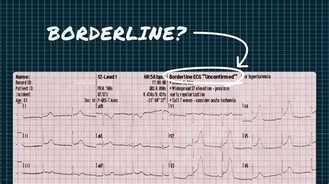
Electrocardiograms (ECGs) are a crucial diagnostic tool in the field of cardiology, providing valuable insights into the heart's electrical activity. While ECG results can be relatively straightforward, some may yield borderline or inconclusive results, leaving both patients and healthcare professionals seeking further clarification. In this article, we will delve into the world of ECG interpretation and explore five signs that may indicate a borderline ECG result.
Understanding ECG Interpretation
Before we dive into the signs of a borderline ECG result, it's essential to understand the basics of ECG interpretation. An ECG measures the electrical activity of the heart, producing a graph that displays the heart's rhythm, rate, and electrical impulses. A typical ECG consists of P, QRS, and T waves, which correspond to the atrial depolarization, ventricular depolarization, and ventricular repolarization, respectively.
Sign 1: Non-Specific ST-T Wave Changes
Non-specific ST-T wave changes are a common finding in ECGs, and they can be a sign of a borderline result. These changes refer to subtle alterations in the ST segment and T wave that do not meet the specific criteria for a particular cardiac condition. For example, a slight ST segment elevation or depression, or a T wave inversion, may be observed. While these changes can be a normal variant, they may also indicate underlying cardiac disease.

Sign 2: Borderline QRS Duration
The QRS complex represents the ventricular depolarization on an ECG. A borderline QRS duration refers to a QRS complex that is slightly prolonged, but not significantly enough to meet the criteria for a bundle branch block or other cardiac conditions. A QRS duration of 100-110 milliseconds is generally considered borderline. This finding may indicate a mild conduction abnormality or a non-specific finding.

Sign 3: Mild Left Ventricular Hypertrophy
Left ventricular hypertrophy (LVH) is a common finding on ECGs, and it can be a sign of various cardiac conditions, including hypertension and cardiomyopathy. Mild LVH is a borderline finding that may not meet the specific criteria for LVH. This can be indicated by a slightly increased QRS voltage or a mild repolarization abnormality.

Sign 4: Non-Specific T Wave Inversions
T wave inversions are a common finding on ECGs, and they can be a sign of various cardiac conditions, including myocardial infarction and cardiomyopathy. Non-specific T wave inversions refer to T wave inversions that do not meet the specific criteria for a particular cardiac condition. This finding may indicate a mild repolarization abnormality or a non-specific finding.

Sign 5: Borderline QT Interval Prolongation
The QT interval represents the time from the beginning of the Q wave to the end of the T wave on an ECG. A borderline QT interval prolongation refers to a QT interval that is slightly prolonged, but not significantly enough to meet the criteria for a QT interval prolongation. This finding may indicate a mild repolarization abnormality or a non-specific finding.

Gallery of ECG Tracings


Frequently Asked Questions
What is an ECG?
+An ECG is a diagnostic test that measures the electrical activity of the heart.
What is a borderline ECG result?
+A borderline ECG result is a finding that does not meet the specific criteria for a particular cardiac condition, but may still indicate a mild abnormality.
What are the signs of a borderline ECG result?
+The signs of a borderline ECG result include non-specific ST-T wave changes, borderline QRS duration, mild left ventricular hypertrophy, non-specific T wave inversions, and borderline QT interval prolongation.
We hope this article has provided valuable insights into the world of ECG interpretation and the signs of a borderline ECG result. Remember to consult with a healthcare professional if you have any concerns about your ECG results.
